“The global graphene market size reached US$ 0.1 Billion in 2022. Looking forward, IMARC Group expects the market to reach US$ 1.54 Billion by 2028, exhibiting a growth rate (CAGR) of 47.80% during 2023-2028.
The growing advancements in the production of semiconductors, rising improvements in various BIOMEDICAL applications, and increasing demand for stronger, lighter, and more durable construction materials are some of the major factors propelling the market.”
— 2022 Graphene Market Report by IMARC
Previous Entries
I — What’s In A Clot
A. Introduction
The precise biological reasons for the pervasive clotting among alleged COVID patients — as well as the jabbed — is still extremely opaque, with a myriad of contradictory explanations. The more honest mainstream researchers will freely admit that “it is not known how abnormal blood clots form in COVID-19 or why they occur even in asymptomatic and convalescent patients.” (Ryu et al.) The physical characteristics, the size, and the systemic nature of the clotting is, dare I say, unprecedented. Even for those who posit that the alleged spike protein is the culprit of this phenomenon, they can not answer why earlier corona viruses did not exhibit these characteristics.
The Regime’s glorified chat bot, ChatGPT, admits as much. I’ll spare you the viral myth making that the chatbot engages in and pare down its answer to what matters:
Yes, spike proteins are not unique to the SARS-CoV-2 virus (which causes COVID-19). Many other viruses, especially those belonging to the Coronaviridae family, have spike proteins…
…spike proteins are common components of various viruses…
Even in this torrent of horse excreta, it is made clear that the “spike protein” is not a unique feature solely attributable to COVID-19.
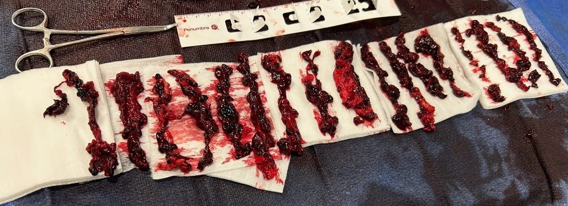
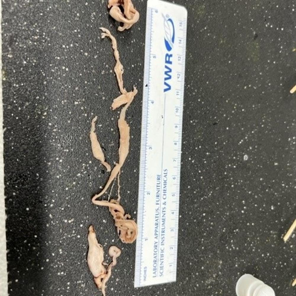
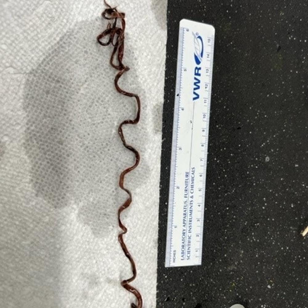
These structures have often been referred to as amyloid fibrous clots in the literature on COVID-19 and “vaccine” injury. Clots should normally contain protein, and they should readily dissolve under hot water. The fibrous structures pulled out of patients — both the living and those who died suddenly — do not exhibit this characteristic. They have been described by rightly concerned embalmers across the world as rubbery in their texture.
How then are the “clots” being formed, and what is causing it amongst both the “positive” cases and asymptomatic cases? Why are these supposed clots not responding to cocktails of high-powered, anti-thrombotic drugs?
Simply put, because they are not clots.
— Clot-like structures harvested from a living patient, May 2023. (Source: Dr. S)
B. Mike Adams Analysis
These Structures exhibit the following Properties.
They are tough, fibrous and resilient, showing material Properties similar to small Rubber Bands.
They consist of many Strands of small, fibrous Strands.
These fibrous Strands (see the very last Photo Set below) show repeating Patterns of scale like Engineering, as if the Body has been programmed to build another Life Form inside the Blood Vessels.
There are strange Crystalline like Structures found on these Clots, exhibiting Transparency and Resistance to normal Gram staining Techniques.
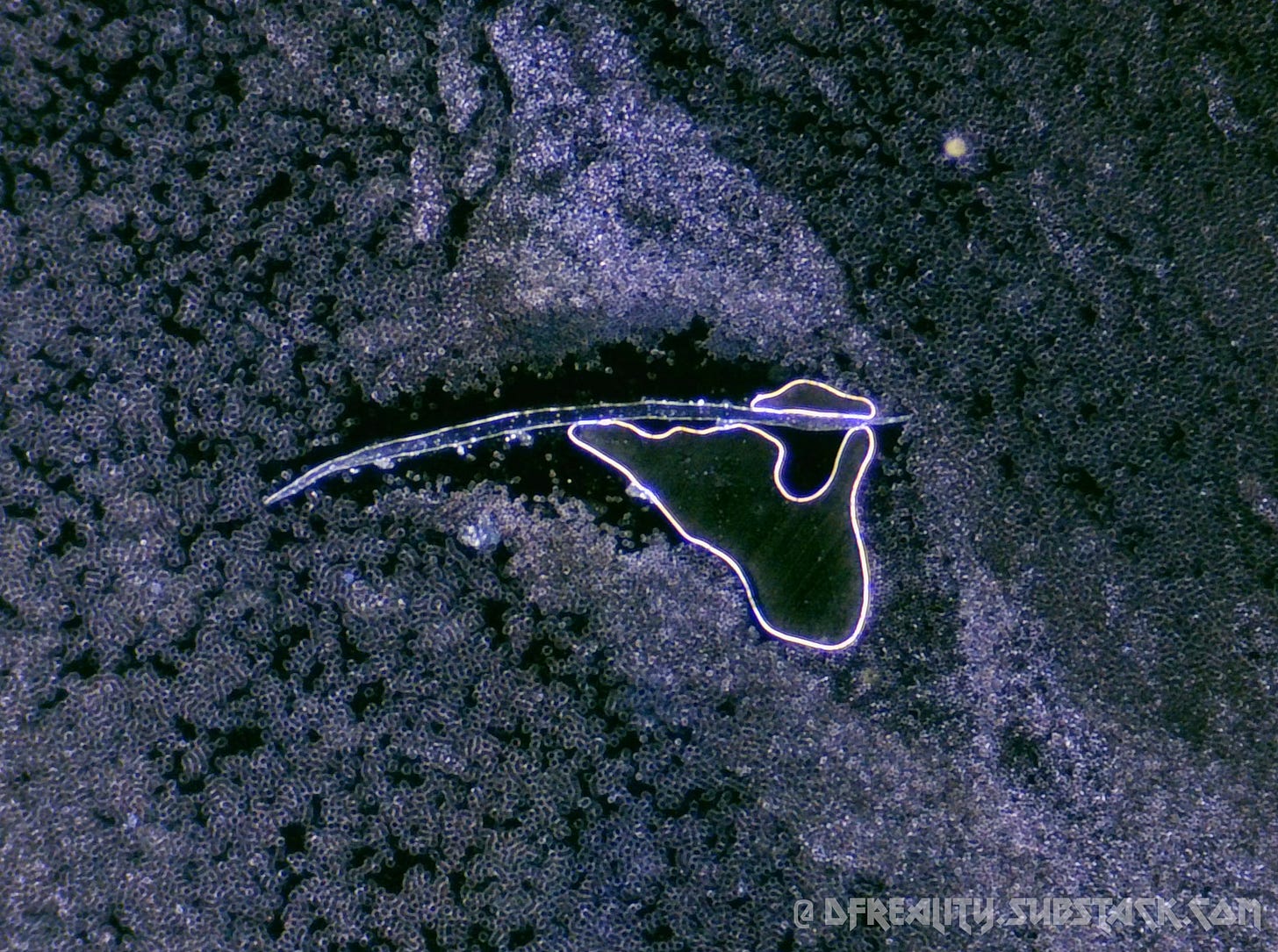
Below, you will find one Example of A Structure that appears to resemble a Silicon like Bio Circuitry or Micro Chip like Structure. We don’t yet know what it is.
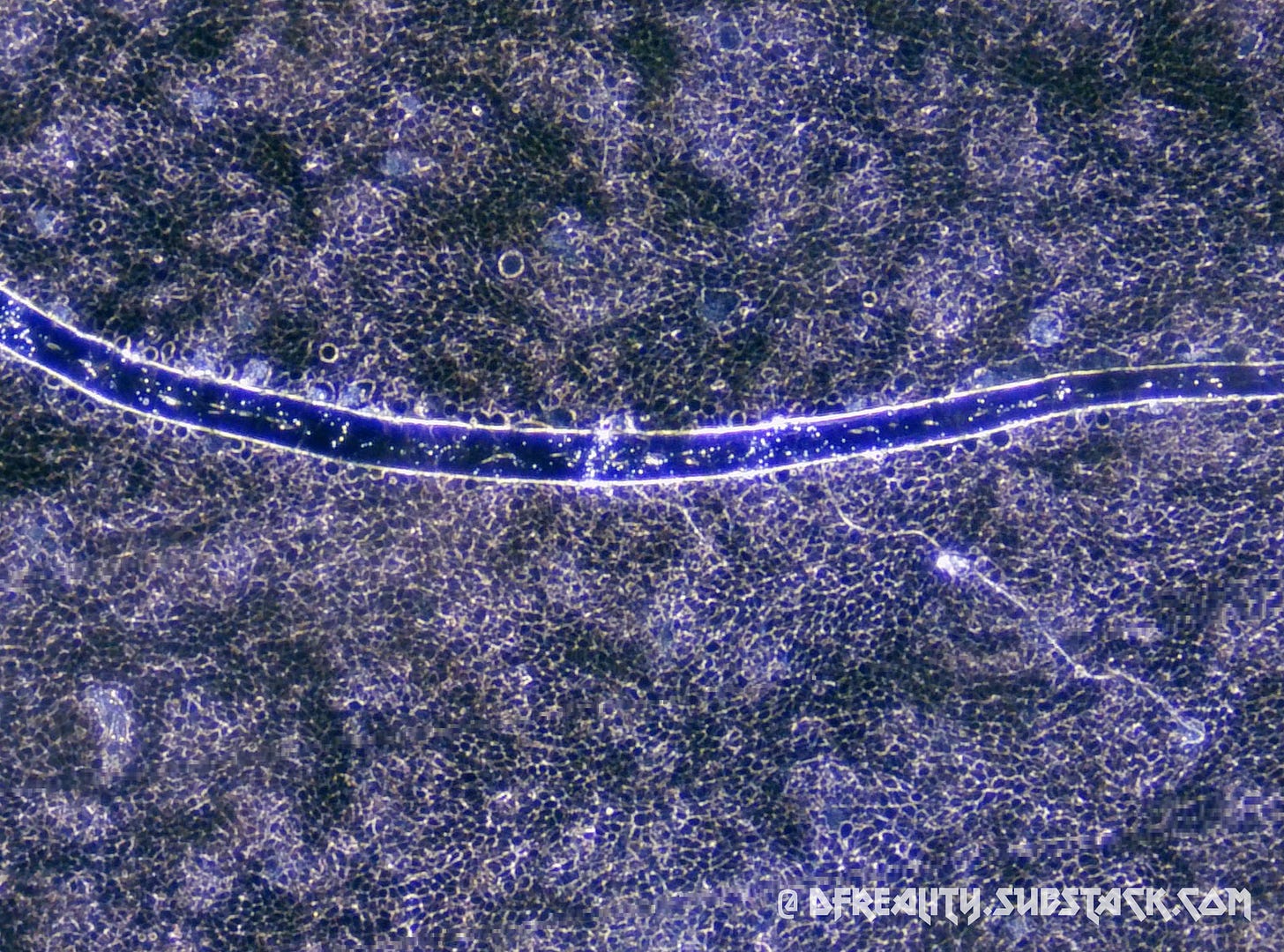
One of the Photo Sets below reveals what appears to be a Bio Circuitry Wire which clearly shows repeating Patterns and Nano Scale Interface Structures that are assembled in A specific Geometry for an unknown Purpose.
Context for the Photos you are about to see.
I received these “Blood Clot” Samples from a reputable Embalmer, Richard Hirschman, who is active in the Field of Embalming and who confirmed these are not Blood Vessels or other Tissues of any Kind. They are Structures that were evacuated from Inside Blood Vessels during embalming Procedures.
I stained these Samples using standard Gram staining Techniques used for Microbiology in order to enhance structural Contrast during Microscopy. One of the Samples below, the more Yellowish Sample, was stained only with Iodine, not any Violet colored Stains.
The Samples were then washed with Ethyl Alcohol and prepared on Slides using standard Tissue Sample Preparation for Microscopy.
Microscope Magnification varies from 20 X to 1500 X, depending on the Photo shown below. Magnifications are indicated with each Photo Set.
— Not Blood Clots, but Structures Extracted from Those who Have Suddenly Died by Mike Adams, Natural News
(20x) (Source: Mike Adams)
(50x) (Source: Mike Adams)
(100x) (Source: Mike Adams)
(200x) (Source: Mike Adams)
(500x) (Source: Mike Adams)
The visuals of these advanced structures and ribbons are staggering in their own right. Chemical analysis of these structures have been analyzed via Inductively coupled plasma mass spectrometry (ICP-MS) in an accredited, audited, inspected and validated laboratory setting. The results make it clear that these structures can simply not be made with human blood due to their chemical composition, with elements that are wildly different than that of normal blood.
C. Dr. Cole Disputes the Findings of Mike Adams
Dr. Ryan Cole founded Cole Diagnostics. His recent report was featured on World Net Daily, Steve Kirsch on Substack, Liberty Counsel, and Just the News. Cole has done multiple interviews since the onset of COVID-19, in which he promoted other alternative means of treating the virus, such as Vitamin D intake, Ivermectin or Hydroxychloroquine. More recently, Cole has been sounding the alarm on what his independent lab in Idaho, the largest in that state, has been noticing when it comes to cancer cases and cancer resurgence since the release of the COVID-19 vaccine.
Now this same lab is reporting the unusual blood clots it examined are primarily made up of protein cells, and this protein production is brought on by spike proteins the body is forced to make, either by infection from the COVID-19 virus, or getting the mRNA vaccine.
Cole believes the greater harm by far seems to be done by the vaccine since the mRNA injection forces the body to produce the spike proteins for a minimum of eight weeks after injection, whereas someone with the virus could halt that production within the body by simply taking Ivermectin or some other early treatment.
In his analysis of these clots, Cole also refers to the studies of South African doctor Resia Pretorius in isolating how the spike protein, whether from COVID or the vaccine, has been a primary cause of clotting and platelet dysfunction.
When asked about his part in supplying Cole Diagnostics with sample blood clots, Hirschmann said he can neither confirm nor deny that Cole’s analysis comes from what he sent. In recent interviews, Cole has acknowledged that other embalmers from other states have found these unusual clots as well and sent samples.
Another lab that has released its findings from the specimens provided by Hirschmann is run by Mike Adams, the founder of Natural News Website. Adams does confirm those samples are from Hirschmann. His report shows highly magnified pictures of these clots and concludes that there is nothing like this ever before found in a decedent who has natural clotting occur from point of death and onwards.
— 'Concerned for humanity': Alabama embalmer discovers strange clots in people since release of COVID vaccine by Andrea Tice
With no chain of custody on where the clots came from, no written reports on how many tests were run, and no revelation of his diagnostic methodologies, Cole’s findings are entirely insufficient — at best. Given Dr. Cole’s self-professed level of expertise in this arena, it is hard not to see his shoddy workmanship and dismissal of the legitimate concerns of nanotechnology as a deceptive attempt at gate-keeping.
II — Not An Accident, But By Design
A. Graphene Is The Culprit
The wound patterns ascribed to COVID-19, Long Covid, or “vaccine” injuries are all readily explained in the scientific and medical literature by nanomaterial exposure: specifically, graphene and graphene derivatives, including hydrogel.
B. Graphene Oxide Causes Thrombosis
Here we report for the first time that atomically thin GO sheets elicited strong aggregatory response in platelets through activation of Src kinases and release of calcium from intracellular stores. Compounding this, intravenous administration of GO was found to induce extensive pulmonary thromboembolism in mice. Prothrombotic character of GO was dependent on surface charge distribution as reduced GO (RGO) was significantly less effective in aggregating platelets.
— Thrombus inducing property of atomically thin graphene oxide sheets by Singh et al.
Carbon aerogels exhibit 3D network, a high porosity structure and high specific surface area, they were synthetized from graphene oxide (GO), whose intrinsic properties make it has great potential in the biomedical applications, such as cancer therapy, tissue engineering, drug delivery, wound healing, biosensing and hemostasis [4–9]. The GO sheets cause strong aggregation in platelets by activating Src kinases and releasing calcium from intracellular stores, which is in a scale comparable to that triggered by thrombin [10]. In addition, activated platelets carry many other hemostatic factors that adhere to the wound surface and trigger the coagulation pathway [9], while graphene aerogels can rapidly absorb plasma, allowing blood cells to accumulate on the surface, further promoting blood coagulation on the wound surface [11]. However, the fatal shortcomings of GO, such as high hemolysis rate [12], potential thrombosis [10], and cytotoxicity [13], limit its application in hemostatic materials.
— Preparation and evaluation of Bletilla striata polysaccharide/graphene oxide composite hemostatic sponge by Chen et al.
C. Graphene Destroys the Immune System, Induces Cytokine Storms, and Causes Inflammation
Under in vitro conditions graphene and GO cause an increased production of pro-inflammatory cytokines, mainly IL-1, IL-6, IL-10 and TNF-a, as a result of the activation of Toll-like receptors in macrophages. Graphene activates apoptosis in macrophages through the TGFbr/Smad/Bcl-2 pathway and also through JNK kinases that are stimulated by an increase of ROS in the cell or through a signal received by Smad proteins. Under in vivo conditions, graphene nanomaterials induce the development of the local inflammatory reaction and the development of granulomas in parenchymal organs.
— The Molecular Influence of Graphene and Graphene Oxide on the Immune System Under In Vitro and In Vivo Conditions by Dudek et al.
D. Hydrogel Mimics Natural Blood Clots
As such, mimicking the functional and material properties of blood clots are a great starting point for development of artificial wound healing materials. From many potential options, we find that hydrogel materials offer an attractive combination of dynamically tunable biochemical functionality, mechanics, and biocompatibility that maps well onto the temporospatial plane of blood clots.
— Synthetic hydrogels as blood clot mimicking wound healing materials by Rausch et al.
Most importantly, the hydrogel can stop high-pressure bleeding from pig carotid arteries with 4~ 5 mm-long incision wounds and from pig hearts with 6 mm diameter cardiac penetration holes.
— A strongly adhesive hemostatic hydrogel for the repair of arterial and heart bleeds by Hong et al.
E. Nanomaterials Are Designed to Mimic “Viruses”
Material scientists have learned much in the last three years of pandemics. On one side, nanomaterial technologies have improved observing viruses [177], mimicking viral strategies used to hijack cells or evade immune responses. Indeed, several distinct viral traits such as size and surface, multivalent ligands, glycosylation, charge, stimuli responsiveness (pH, ROS and enzymatic triggers), and the sequential interactions with cellular receptors might help creating nanomaterials with better pharmacokinetics and delivery advantages [178].
Materials science has also created thousands of new solutions to quickly respond to pandemic diseases by creating devices that detect viral proteins, point-of-care diagnosis and monitoring methods, and treatment and vaccination strategies [172].
…Among nanomaterials, [carbon-based materials] have been synthesized in a variety of shape, sizes and functionalized and tested in many different biological systems in the last 15–20 years.
— Nanotechnology in the COVID-19 era: Carbon-based nanomaterials as a promising solution by Papi, De Spirito, Palmieri
F. Summary
Graphene oxide introduced into the body induces thrombogenicity and the formation of blood clots.
When graphene oxide is present within the body, it gives rise to post-inflammatory syndrome, systemic inflammation, and inflammation affecting multiple organs.
Graphene oxide, exceeding the levels of the body's natural antioxidant reserve — glutathione — results in immune system disturbance, immune system collapse, and the onset of cytokine storms.
Upon inhalation, graphene oxide disperses uniformly throughout the alveolar tract, provoking bilateral pneumonias. (Zhang et. al.)
Inhaled graphene oxide triggers inflammation in the mucous membranes, leading to the loss of taste and smell, medically known as anosmia.
When it accumulates at the neuronal level, it precipitates neurodegeneration, essentially inducing neurological COVID-19 (a.k.a. Brain Fog).
In summary, graphene oxide closely mimics the symptoms and wound pattern of COVID-19, resembling the characteristics of the purported SARS-CoV-2 in mainstream accounts.
III — Graphene: A Current and Growing Threat
A. Graphene Is Already Being Used in Biosensors
Many of these new realities are becoming possible through the design and use of nanosensors, which can “identify specific cells at the molecular level to deliver medicine and monitor the development of particular places in the body by measuring physical characteristics such as volume, concentration, movement and speed, gravitational, electric, magnetic forces, pressure, temperature, etc.” [158].
Nanosensors are sensing devices made from or with engineered nanomaterials and can be manufactured with the following:
• Nanoscale wires.
• Carbon-based nanomaterials (carbon nanotubes [CNTs] and graphene [GR]).
• Thin films.
• Metal and metal oxide [nanoparticles].
• Nanoscale polymers.
• Nanobiomaterials.
Two of the most important and currently used and researched materials for nanosensors are carbonbased nanomaterials and metal NPs. According to Javaid et al. [158], carbon-based nanomaterials offer improved conductivity, stability, lower costs, and ease of surface functionalization compared with traditional sensor materials. Common carbon nanomaterials used in sensing include CNTs and GR, which have been evaluated for use in chemiresistive sensing. Some recent applications of carbon-based GR nanomaterials for biosensors are found in Table 6-1.
Metal NPs are another commonly used type of nanomaterial of biosensing. These types of sensors can often be more sensitive and selective due to the ability to use custom signal amplifications [158]. Commonly explored metals for this purpose include cobalt, Cu, Au, palladium (Pd), platinum (Pt), Ag, and others.
— Nanotechnology Applications for Combat Casualty Care by Nichols, Shelby, & Milonas. August 2022.
— Table 6-1. Carbon-Based Nanomaterial Applications Used in Nanobiosensing (Source: Bardhan et al.)
Graphene has been a new star for application in various fields, such as catalysis [1], energy storage [2] and electrochemical biosensors [3] since its discovery in 2004. The applications of graphene in biosensors are focused on several fields. First, graphene or functionalized graphene has been used to modify the substrate such as GCE [4], [5], [6], ITO device [7], and quartz chip [8] because of their excellent conductivity and high chemical stability. Then graphene or graphene oxide can also act as a quencher to quench the fluorescence of illuminant for its unique electronic properties [9], [10], [11]. Li and his coworkers have realized real-time target monitoring in living cells by graphene oxide [10]. Last but not the least, graphene has high surface area; more and more graphene-based nanocomposites with different kinds of functions as enhanced sensing material have been reported [7], [12], [13].
— Label-free aptamer biosensor for thrombin detection based on functionalized graphene nanocomposites by Wang et al.
B. Graphene Nanoribbons: Precursors to Emboli
— Blood vessel size comparisons. (Source: Fundamentals of Nursing, Pearson Publishing.)
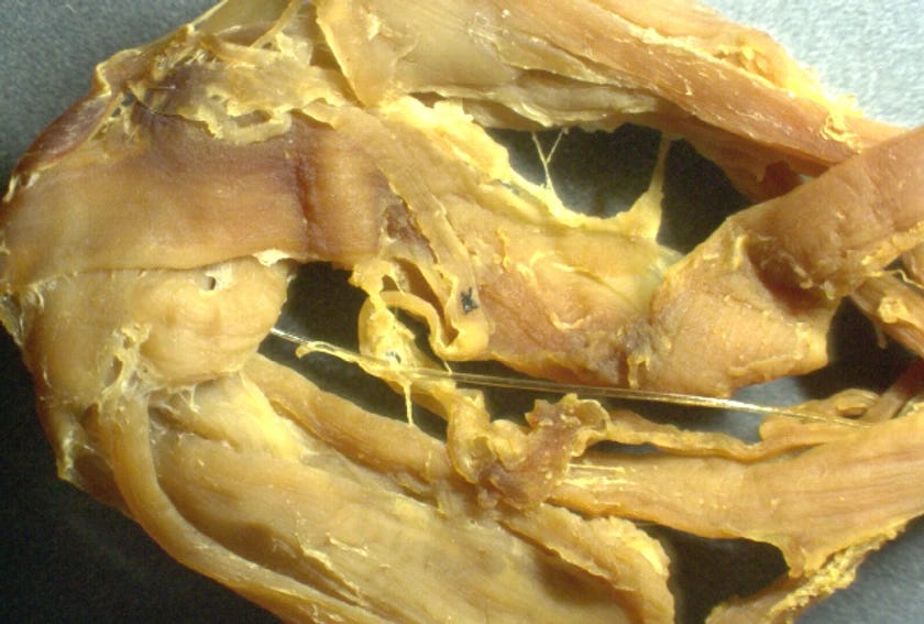
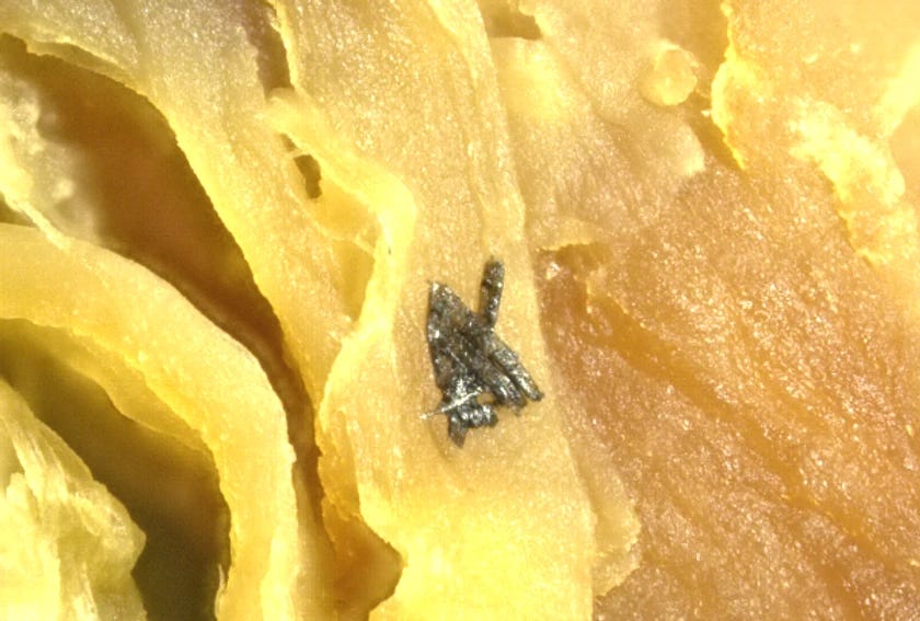
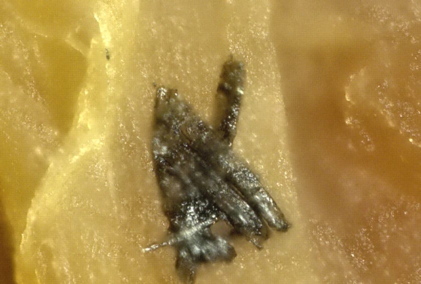
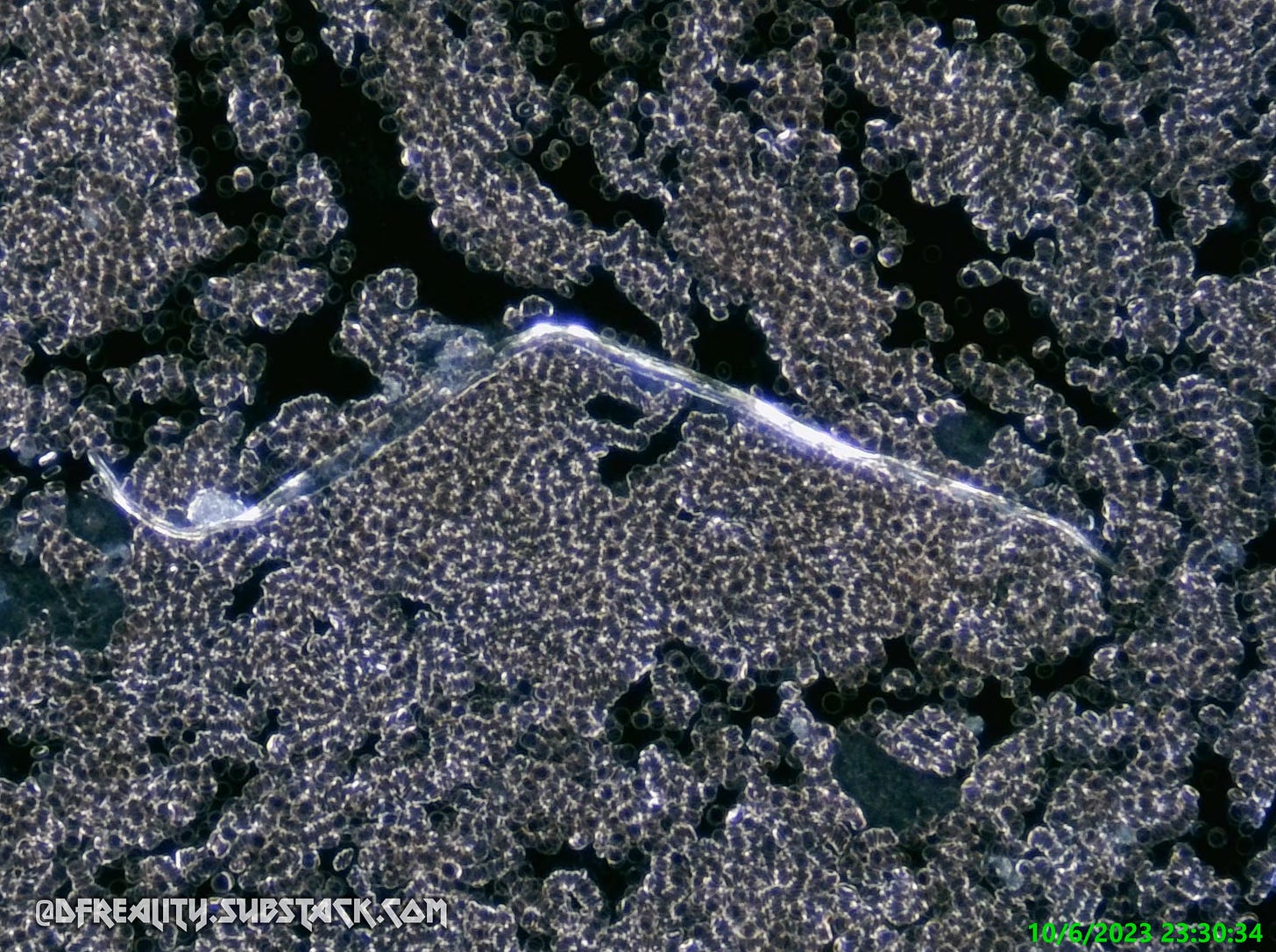
For scale, the tiny circles in these pictures from previously collected samples are blood cells, measuring just 7-10 μm (micrometers) wide. Our findings clearly demonstrate the risk for capillary, venule, and arteriole blockages from graphene ribbons as they are traveling through the tiniest parts of our circulatory system.
C. Conclusions
The prevailing narrative surrounding COVID-19 and its treatment through vaccines has left us ensnared in a web of obfuscation and deceit, spun by those who claim to act in our best interests. As we delve deeper and deeper into the murky depths of this grand deception, it becomes alarmingly evident that the essence of our struggle extends far beyond the invisible battlefield of premeditated pandemics and venomous vaccines:
It is a battle for truth, transparency, and the preservation of human autonomy.
As we continue to analyze the evidence, the tentacles of graphene's insidious influence become unmistakable. The so-called "spike induced clots" at the heart of this enigma are but a smokescreen, concealing the true malevolence at play. We find ourselves confronted not by mere happenstance, but by a calculated and meticulous design. It is no mere accident that materials of this nature have found their way into the bulk of humanity. The much-touted "spike protein" is but one piece of this intricate puzzle, an illusory danger carefully woven into the fabric of nanotechnology; concealed within the core of a graphene laden conspiracy. (See Chaff & Countermeasures for a further exploration of this subject.)
The forces at work here are not bound by morals or ethics, discarding such quaint trappings long ago. Graphene Oxide’s role in mimicking viral strategies; introducing thrombogenicity; decimating the immune system; inducing cytokine storms; and sowing inflammation is revealed as the linchpin of this scheme. The insidious use of hydrogels to replicate the attributes of natural blood clots further underscores the deliberate artifice at play. Thusly, it allows the malevolent orchestrators behind this agenda to continue to maim and manipulate our very biology.
The stakes are far too high for us to continue to be passive observers.
It is incumbent upon us to awaken from this nightmare of misinformation and complacency.
We must rally against the darkness that seeks to engulf us, persevering so that the light of Christ’s Truth prevails.
"We are now at a CROSSROADS. One path will take us to a better world: more inclusive, more equitable and more respectful of Mother Nature. The other will take us to a world that resembles the one we just left behind – but worse and constantly dogged by nasty surprises. We must therefore get it right.
The looming challenges could be more consequential than we have until now chosen to imagine, but our capacity to reset could also be greater than we had previously dared to hope.”
― Klaus Schwab, COVID-19: The Great Reset







The only only thing I would caution here concerns the subject of Mike Adams. I have had absolutely no doubt he is controlled opposition since about 2017, long before the Convid Op. He may be telling the truth about this but is still not to be trusted in my opinion. He came straight out of Stratfor in Austin, Texas, just like Alex Jones. Alex was the big project by Stratfor to create a replacement for Bill Cooper who was controlled. When Alex took off they murdered Cooper. My guess is Adams was created specifically to have a specialty, Convid version of Alex. Brighteon has become a huge nest of these kinds of operatives. All of the Lucis Trust, shill doctors are on there. Northrup, Tenpenny, Madej, etc.
Great source. There are still trolls/bots/morons, who say graphene doesn't even exist...